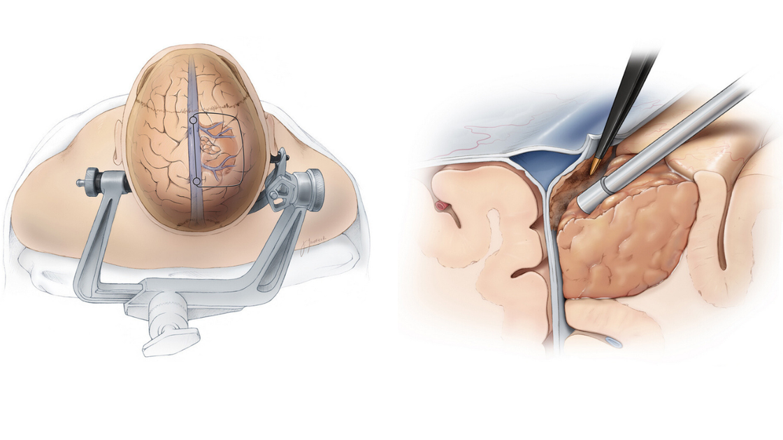
Is Brain Tumour Surgery Risky? What Patients Need to Know Before Making a Decision
Related Hospitals
Discover hospitals and medical centers related to this topic for quality healthcare services.
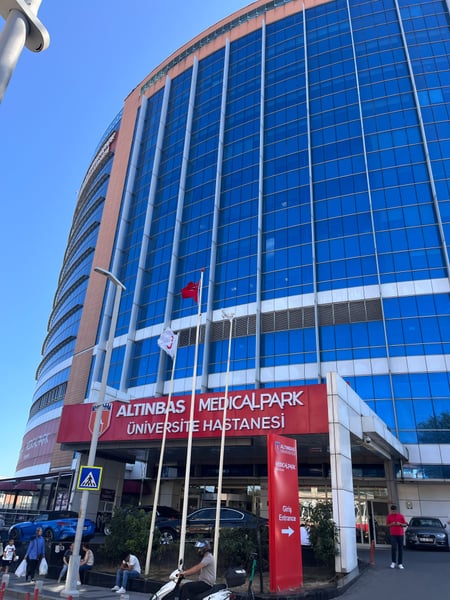
Medical Park Bahcelievler Hospital
Medical Park Bahcelievler Hospital is a 242-bed JCI-accredited hospital in Istanbul, established in 2007. Spread across 33,000 square meters and 19 fl...
Accreditations
Facilities
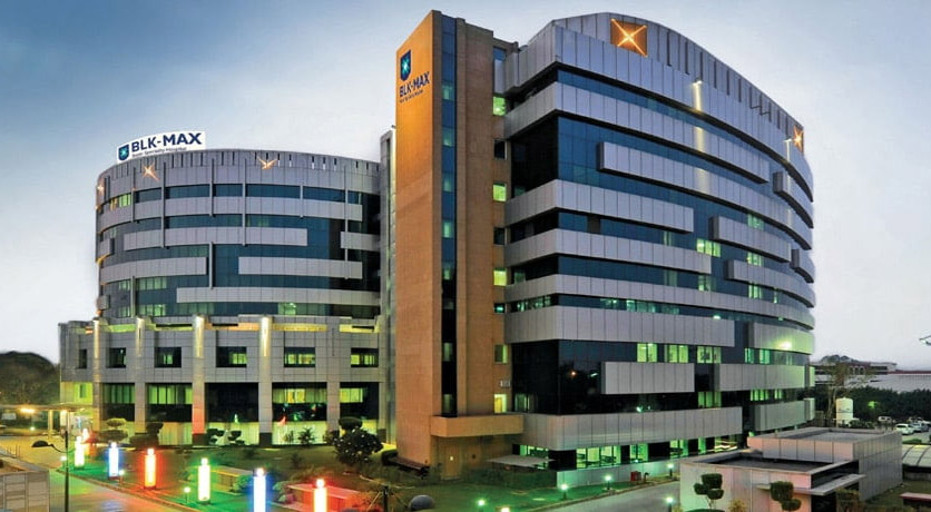
BLK-Max Super Speciality Hospital, New Delhi
BLK-Max Super Speciality Hospital in New Delhi is one of India's premier healthcare institutions, offering 650 beds, 22 advanced operation theatres, a...
Accreditations

Facilities
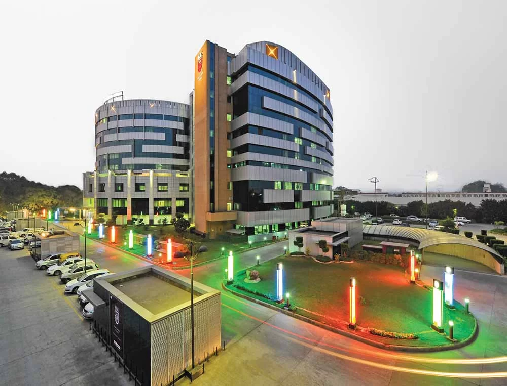
Fortis Memorial Research Institute (FMRI), Gurgaon
Fortis Memorial Research Institute (FMRI), Gurgaon, is a world-class multi-specialty hospital established in 2013. The hospital offers 330 beds, 15 op...
Accreditations
Facilities

Artemis Hospital, Gurgaon
Artemis Hospital, Gurgaon, is a JCI accredited multispecialty hospital that was established in 2007. It offers 750+ beds and world -class infrastructu...
Accreditations

Facilities
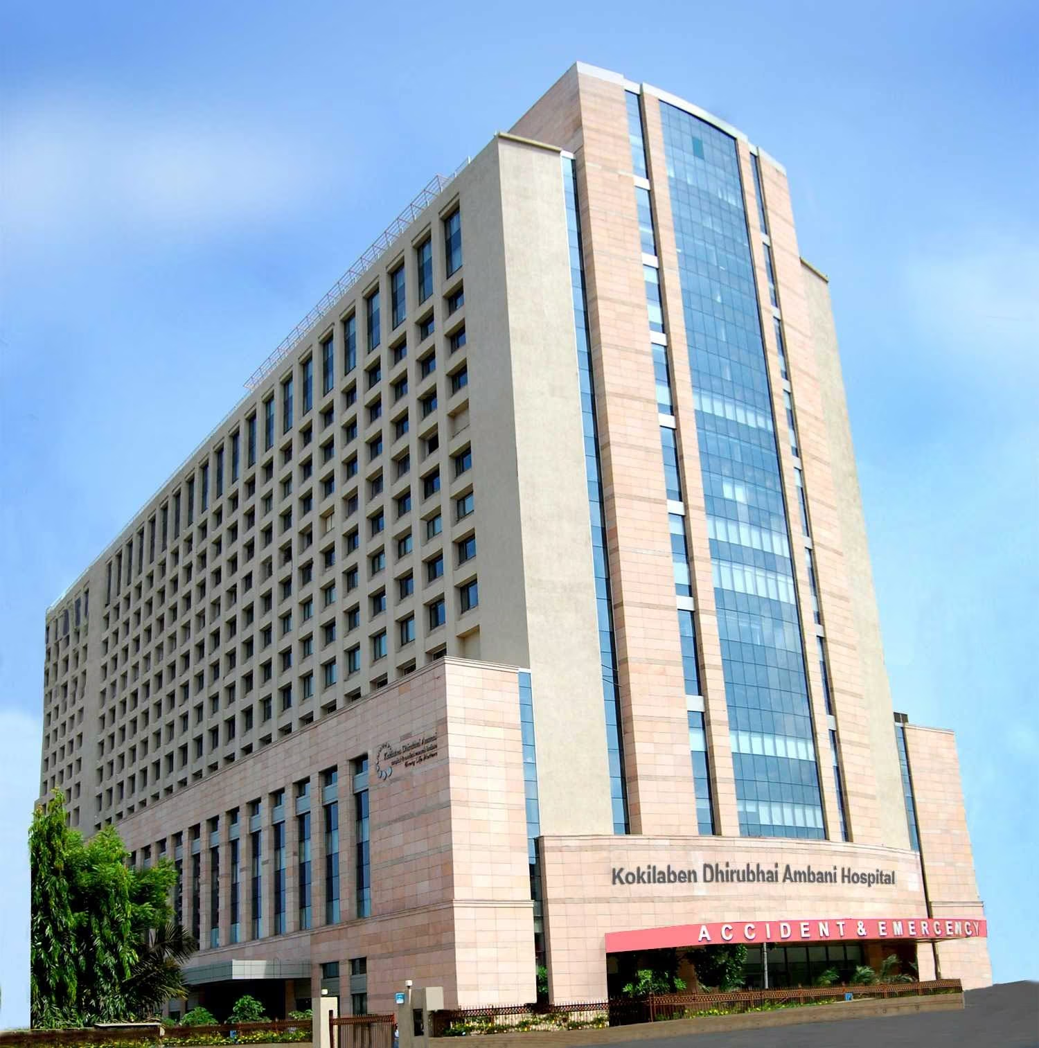
Kokilaben Dhirubhai Ambani Hospital, Mumbai
Kokilaben Dhirubhai Ambani Hospital, Mumbai, is a JCI, NABH, NABL, and CAP-accredited quaternary care hospital established in 2009. With 750 beds, 180...
Accreditations
Facilities
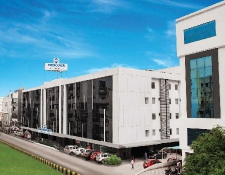
Medicover Hospitals, Hitech City, Hyderabad
Medicover Hospitals, Hitech City, Hyderabad, is a 400-bed NABH-accredited super-specialty hospital established in 2011. It is part of Medicover, a glo...
Accreditations
Facilities
Related Doctors
Connect with experienced doctors and medical specialists in this field.

Dr. Wichit Arpornwirat
Consultant

Dr. Prasan Kachonrattanadet
Consultant

Dr. Suporn Chuncharunee
Associate Professor

Dr. Ekaphop Sirachainan
Associate Professor

Dr. Ittichai Sakarunchai
Associate Professor

Dr. Sombat Muengtaweepongsa
Associate Professor
Related Articles
Explore more articles and insights on similar health topics.

How Multidisciplinary Teams Improve Patient Outcomes

Why Hospital Accreditation Matters in Medical Tourism

Managing Pain After Spine Surgery

How Long Does Recovery Take After Spine Surgery?
Post-Transplant Care for International Patients

Organ Transplant Surgery: Risks, Benefits, and Long-Term Survival

Our website uses cookies. By clicking on accept you give your consent to the use of cookies as per our Privacy Policy.